Why Your Pain Doesn’t Show Up on Imaging
Many people who experience chronic pain find themselves frustrated after imaging tests like MRIs, X-rays, or CT scans come back “clear” or “normal.” They know their pain is real, but the images often don’t reveal anything conclusive. Medical gaslighting anyone?
This disconnect between what we feel and what imaging shows can be confusing, but it often boils down to one simple reason: restricted fascia.
Fascia: The Missing Piece
Fascia is a connective tissue that supports every muscle, nerve, organ, and blood vessel in our body. It is a 3D web-like structure that connects all parts of the body, offering both mobility and stability.
Healthy fascia glides freely, is flexible, and resilient, allowing you to move without restriction and allows vital processes like the flow of blood, lymphatic fluid, and nerve conduction to occur.
However, when fascia becomes stuck down or restricted due to factors like injury, repetitive strain, emotional stress, or lack of movement, it can cause significant pain.
Unfortunately, traditional imaging methods do not detect fascial restrictions.
Fascia doesn’t show up clearly on MRIs or X-rays, which are primarily designed to look at bones, organs, and major structures.
Even soft-tissue scans like Ultrasound and CT-Scan often miss the subtle changes in fascia that could be causing chronic discomfort.
As a result, people with fascial restrictions may leave medical appointments with little explanation for their pain.
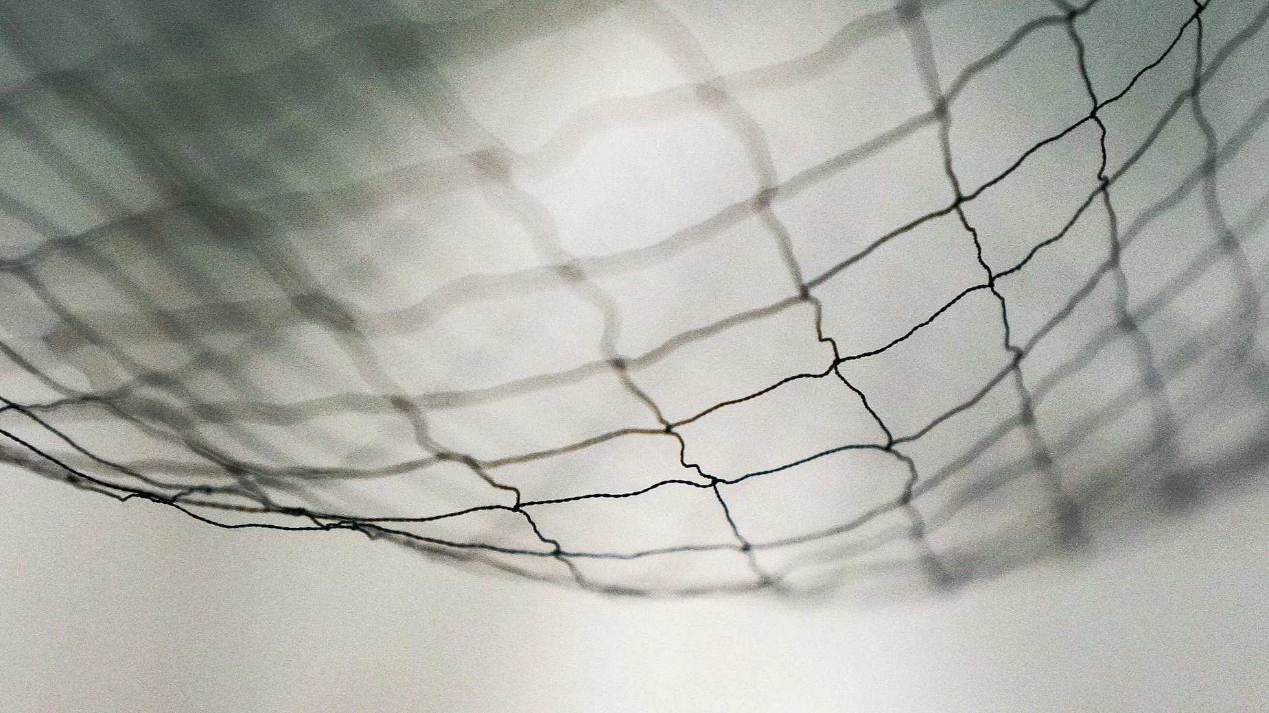
Fascia exists as a 3D web of connective tissue
Why Traditional Imaging Often Misses the Mark
1. Focus on Structure, Not Function: Most imaging techniques are designed to identify structural abnormalities, such as broken bones or torn ligaments. They are not as sensitive to functional issues, like restricted or immobile fascia, which can create tension and inflammation but may not appear damaged or different on an MRI or X-ray.
2. Lack of Specificity for Soft Tissue: While MRIs can sometimes detect soft tissue issues, they generally don’t capture the subtle changes within fascia. Fascia is thin and dynamic, and its condition is best understood through its ability to move and adapt, not just how it looks in a static image.
3. Missed Connections Between Symptoms and the Fascia Network: Fascial restrictions don’t always cause pain at the source. Pain in one part of the body may be caused by tension or restriction in a completely different area due to fascia’s interconnected structure. This is often missed in localized imaging studies.
Still want answers for your pain?
A Myofascial Release Therapist trained in the John Barnes method is expertly capable of finding areas of fascial restriction in the body based on a combination of reported symptoms, postural analysis, and feelings of heat, vibration, or tenderness on palpation.
Typically fascial restrictions are also areas of inflammation and the body often expends a lot of heat energy at the site of restriction in an attempt to heal.
For this reason, Thermography, a type of infrared imaging, offers a promising way to visualize the effects of fascial restrictions.
Unlike traditional imaging, thermography detects heat patterns on the body’s surface, which can indicate inflammation or unusual temperature changes.
These heat patterns often correlate with areas of fascial tension or restrictions, providing a visual clue to the origin of pain.
Key advantages of thermographic imaging include:
1. Detecting Inflammation and Circulatory Changes: Thermography is highly sensitive to changes in blood flow and inflammation. When fascia is restricted, blood flow may be reduced or re-routed, creating “hot spots” that thermography can detect. These spots often correlate with areas of pain and tension that other imaging techniques overlook.
2. Non-Invasive and Radiation-Free: Thermography is completely non-invasive and does not expose patients to radiation, making it a safe alternative for people who need regular monitoring or those sensitive to other imaging techniques.
3. Early Detection: Thermography can often detect subtle physiological changes before structural changes occur. This early insight is especially helpful for chronic pain sufferers, as it can catch issues that might not show up on structural imaging for years.
Examples of Thermographic Images
Understanding Fascia is Key for Pain Relief
When pain doesn’t show up on traditional imaging, it’s easy to feel disheartened.
But understanding the role of fascia can be empowering.
Restricted fascia doesn’t just cause pain in one spot; it can affect the body as a whole, creating patterns of discomfort that radiate far beyond the origin.
Knowing that fascia can be the cause of pain also opens up a new world of treatment possibilities, such as sustained-pressure myofascial release therapy, which focuses on gently and effectively releasing fascial restrictions to restore function and reduce pain.
At the intersection of myofascial work and thermography, we find a comprehensive approach to managing and relieving pain that is often “invisible” to traditional scans. This approach allows us to address the root causes of pain rather than simply managing symptoms.
Moving Forward with a New Perspective
If you’ve been told your imaging results are “normal” but you’re still in pain, consider looking beyond traditional scans.
Thermographic imaging combined with myofascial release therapy may offer the clarity and relief you need.
By addressing the fascia network, we can better understand, visualize, and relieve the source of your pain, allowing you to regain freedom and movement in your life.
Embracing the role of fascia and exploring advanced imaging options like thermography may be the key to finding relief where traditional methods fall short.


